During cell division the mother cell physically splits into two daughter cells. A contractile actin-myosin ring assembles underneath the plasma membrane when the chromosomes segregate in anaphase. Contractile ring constriction results in the ingression of the cleavage furrow and eventually in the formation of the two daughter cells. The position of the contractile ring is determined by two signals from the mitotic spindle. A stimulatory signal at the cell equator promotes the activation of the small GTPase RhoA in a narrow zone. In parallel, an inhibitory signal from the astral microtubules inhibits contractile ring assembly at the cell poles.
The stimulatory signal depends on the guanine nucleotide exchange factor (GEF) Ect2, which activates RhoA at the cell equator. Our work in human tissue culture cells has uncovered how Ect2 activity is spatially and temporally controlled by its three BRCT domains, which are located at the N-terminus (Schneid et al., Cell Reports 2021)
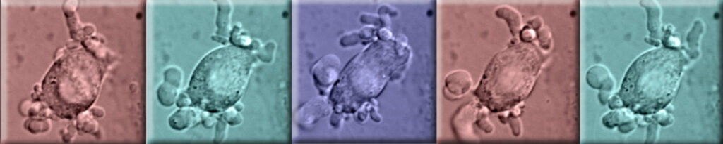
Microscopic images of a human cell during mitosis with hypermembrane blebbing due to uncontrolled Ect2 activity.
The inhibitory signal at the cell poles depends on the dynamic microtubules generated at the centrosomes. Our laboratory discovered the first two molecular components of the inhibitory signal in Caenorhabditiselegans. We demonstrated that the activation of Aurora A kinase by its activator TXPL-1 promotes the removal of contractile ring components from the cell poles during anaphase (Mangal et al. Journal of Cell Biology 2018)
Publications
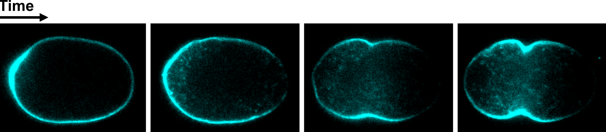
Wild type C. elegans one-cell embryo at different time points during anaphase expressing a F-actin marker (LifeAct-Cyan). F-actin is removed from the cell poles over time.
Cell division is highly dynamic and to study and manipulate it one requires tools with high spatial and temporal precision. In collaboration of the group of Henry Dube we developed a light-controllable proteasome inhibitor (Cage-MG132), which allows us to manipulate cell division with blue light. Using blue light pulse of 5 min we could either arrest cancer cells during the cell cycle or induce cell death (Uhl et al., Angewandte Chemie 2021)
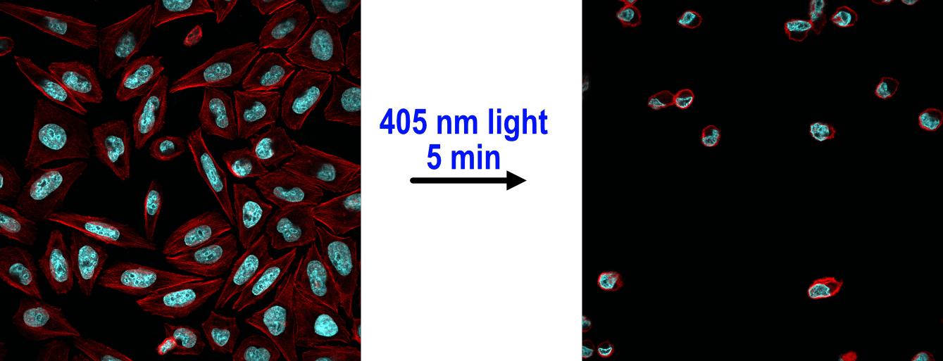
Human cancer cells (HeLa) were incubated with Cage-MG132 and treated with a 5 min pulse of 405 nm light. After 16 hours cells were stained for DNA (blue) and F-actin (red). The light pulse induces cell death in the Cage-MG132-treated (left image) but not the control cells (right image).